Calculate relative wall thickness (RWT) from posterior wall thickness and LV end-diastolic diameter, then classify left ventricular geometry.
Educational use only; not medical advice or a diagnosis. LV geometry labels and cutoffs can vary by lab and must be interpreted by a clinician using a complete echocardiogram and clinical context. If you have chest pain, fainting, shortness of breath, palpitations, or known heart disease, seek urgent care or contact a clinician.
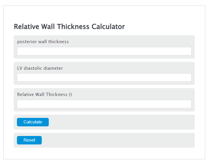
Relative Wall Thickness Formula
Variables:
- RWT is the Relative Wall Thickness (unitless)
- PWT is the posterior wall thickness
- LVD is the LV diastolic diameter
To calculate Relative Wall Thickness, divide the posterior wall thickness by the LV diastolic diameter, then multiply by 2.
Clinical guidance (cutoffs & methods)
Commonly used cutoffs (for example, RWT 0.42 and sex-specific LV mass index thresholds) and the LV mass calculation approach can differ by echocardiography lab, patient population, and indexing method (e.g., BSA vs height-based indexing). For the most widely used conventions, check the American Society of Echocardiography (ASE) / European Association of Cardiovascular Imaging (EACVI) “chamber quantification” guideline documents and your local echo lab’s reporting standards.
How to Calculate Relative Wall Thickness?
The following steps outline how to calculate the Relative Wall Thickness.
- First, determine the posterior wall thickness.
- Next, determine the LV diastolic diameter.
- Next, gather the formula from above = RWT = 2 * PWT / LVD.
- Finally, calculate the Relative Wall Thickness.
- After inserting the variables and calculating the result, check your answer with the calculator above.
Example Problem :
Use the following variables as an example problem to test your knowledge.
posterior wall thickness = 0.78 cm
LV diastolic diameter = 4.0 cm
FAQs
What is the significance of measuring Relative Wall Thickness (RWT)?
Measuring RWT is crucial for assessing the geometry of the left ventricle, which can provide valuable insights into cardiac health. It helps in identifying patterns of remodeling or hypertrophy when interpreted alongside other echocardiogram measurements.
Can Relative Wall Thickness be used to diagnose heart conditions?
RWT helps classify LV geometry (e.g., concentric remodeling/hypertrophy vs eccentric hypertrophy) and is interpreted alongside LV mass index and the full echocardiogram report. It is not sufficient to diagnose specific cardiomyopathies such as hypertrophic cardiomyopathy (HCM).
What are the normal values for Relative Wall Thickness?
A commonly used cutoff is 0.42: RWT ≤ 0.42 is generally considered not increased (normal), and RWT > 0.42 is considered increased. Interpretation of LV geometry (normal geometry vs concentric remodeling/hypertrophy vs eccentric hypertrophy) depends on LV mass index in addition to RWT.
How often should Relative Wall Thickness be measured?
RWT is typically reported when an echocardiogram is performed for a clinical reason. The need for repeat echocardiograms depends on symptoms, diagnoses, and clinician guidance.